


What type of skin hyperpigmentation do I have?
Author
Written by one of our askINKEY skincare advisors
Published
16 June, 2022
Dark spots, patches, and uneven skin tone are among the most searched skincare concerns in the world - but not all of them are the same condition, and treating them as if they are is one of the most common reasons results fall short. This guide exists to solve one specific problem: helping you identify which type of hyperpigmentation you have, based on how it looks, where it appears on your skin, and what triggered it.
The four types covered in this guide are post-inflammatory hyperpigmentation (PIH), melasma, sunspots (solar lentigines), and freckles (ephelides). Each has a distinct appearance, a distinct cause, and a distinct set of visual characteristics that make identification possible without a dermatologist's appointment - though one is always a good idea if you are unsure.
This guide does not cover full treatment routines, ingredient science, or how to build an AM and PM skincare routine. That territory belongs to our complete hyperpigmentation guide, which covers everything from how melanin overproduction works to the most effective ingredients and how to use them. What you will find here are identification checklists, visual descriptors, key comparisons, and type-specific product starting points. If you are specifically dealing with marks left behind after acne, our acne scars guide goes deeper on PIH and post-blemish treatment.
Identification checklists for each type appear in Section 3. Type-specific product callouts are in Section 6. A quick diagnostic summary pulling together all four types is in Section 7.
Products for Hyperpigmentation - Quick Reference
Before getting into identification, here are the products most commonly used to address hyperpigmentation - whichever type you have:
- Tranexamic Acid Serum - $19 - Our hero dark spot serum. AM and PM. All skin types. → How long does Tranexamic Acid take to work?
- 15% Vitamin C + EGF Serum - $20 - Brightening and antioxidant protection. AM. → What is Vitamin C?
- Niacinamide Serum - $13 - Tone-evening and oil control. AM and/or PM. → What is Niacinamide?
- Glycolic Acid Toner - $18 - Exfoliating toner for faster cell turnover. PM only, 2-3x per week.
- Starter Retinol - $15 - Overnight skin renewal. PM only. → What is Retinol?
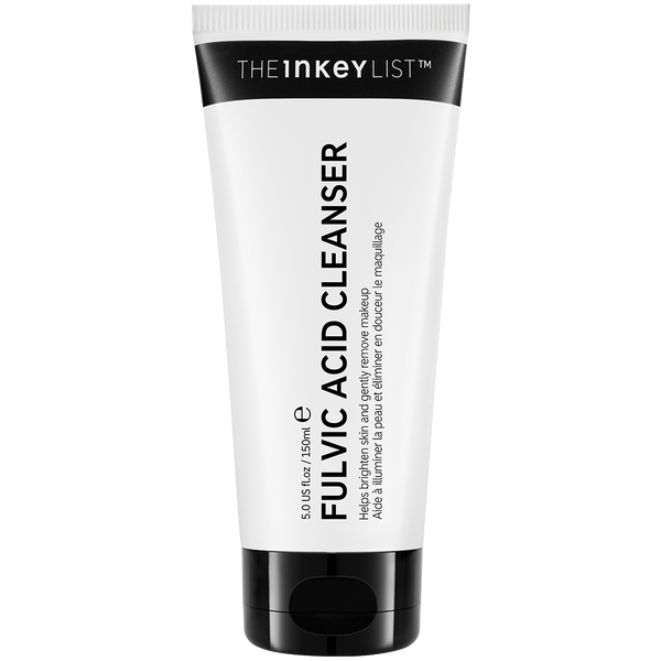
- Fulvic Acid Cleanser - $15 - Brightening cleanser with antioxidant action.
- 10% Azelaic Acid Serum - $20 - Anti-inflammatory and pigment-targeting. AM and/or PM. → What is Azelaic Acid?
- SPF - Daily broad-spectrum sun protection. Essential for all types. → What is SPF? | Why SPF is non-negotiable for hyperpigmentation
Now, to the identification. The four types of hyperpigmentation differ in how they look, where they appear, who gets them, and what causes them. Here is how to tell them apart.
Why Identifying Your Type of Hyperpigmentation Matters
Hyperpigmentation is a broad umbrella term. It describes any area of skin that appears darker than the surrounding tone - caused, in every case, by an overproduction of melanin, the pigment responsible for skin, hair, and eye color. That shared mechanism might suggest that all dark spots are more or less the same problem. They are not.
The four main types of hyperpigmentation - PIH, melasma, sunspots, and freckles - share the same underlying biology but differ significantly in what triggers them, where they appear, how they behave across seasons, and how they respond to treatment. A product or routine that is highly effective for post-inflammatory marks may have limited impact on hormonally driven melasma. An approach designed for melasma may be unnecessary for someone with genetically predisposed freckles. Starting treatment before identifying your type is, at best, inefficient - and at worst, it can make certain types worse.
This blog is the diagnostic companion to our complete hyperpigmentation guide. The pillar covers the full science, the best ingredients, and how to build a routine. This blog focuses entirely on one question: which type do you have?
The four main types of hyperpigmentation are post-inflammatory hyperpigmentation (PIH), melasma, sunspots, and freckles. Here is a detailed breakdown of each.
The Four Types of Hyperpigmentation - How to Identify Each One
Each type below follows the same format: what it looks like, where it appears, what causes it, who is most affected, how skin tone influences its appearance, and a clear identification checklist. Read through each one and note which characteristics match what you are seeing on your skin.
Post-Inflammatory Hyperpigmentation (PIH) - Identifying Dark Marks After Acne
Post-inflammatory hyperpigmentation is exactly what its name describes - pigmentation that develops as a direct response to inflammation. When skin experiences trauma, from acne, a burn, an eczema flare, an ingrown hair, or even aggressive product use, melanocytes (the cells that produce melanin) respond by overproducing pigment. That excess pigment is deposited in the skin and remains visible long after the original inflammation has resolved.
What it looks like
PIH presents as flat, dark marks. The color range is wide: in lighter skin tones, the marks may appear pink, red, or light brown. In medium to deeper skin tones, PIH is typically dark brown or gray-brown, and the contrast with surrounding skin is more pronounced. The marks themselves are completely flat - no raised texture, no rough surface. Running a finger across a PIH mark should feel identical to running a finger across the surrounding skin. The skin's collagen structure is intact; the discoloration is purely a pigmentation issue, not a structural one.
The shape and distribution of PIH marks are irregular and asymmetrical. They appear wherever individual blemishes or areas of inflammation previously were - on the cheeks, jawline, chin, and forehead in acne-prone skin, or anywhere on the face and body where trauma occurred. Unlike melasma, they do not follow a bilateral or symmetrical pattern.
What causes it
The most common triggers for PIH include acne and breakouts, ingrown hairs, eczema and dermatitis flares, burns, cuts and abrasions, skin trauma from procedures, and damage caused by picking, squeezing, or using products that are too harsh for the skin. Any inflammatory response in the skin can produce PIH, which is why it is such a widespread concern.
Who is most affected
PIH affects all skin tones, but it is significantly more common and more pronounced in medium to deeper skin tones. This is because melanocytes are more active at baseline in higher-Fitzpatrick skin, meaning the inflammatory pigmentation response is stronger and the resulting marks are darker and can persist for longer.
Distinguishing PIH from structural scarring and from PIE
Two common points of confusion are worth addressing directly. First, PIH is not the same as a textured acne scar. If the mark is indented (atrophic) or raised (hypertrophic), that is structural scarring involving collagen - a different concern. PIH leaves the surface perfectly smooth.
Second, PIH is frequently confused with PIE - post-inflammatory erythema. PIE presents as pink or red marks and is caused by dilated or damaged blood vessels, not by excess melanin. It responds to different ingredients. The glass test is the most reliable way to distinguish between the two: press a clean, clear glass firmly against the mark. If the color fades or disappears under pressure, it is PIE (vascular). If the color remains visible, it is PIH (melanin-based).
Identification Checklist - Post-Inflammatory Hyperpigmentation
- The mark appeared after acne, a breakout, or an area of inflammation
- It is completely flat - no bump or indentation when you run a finger across it
- It sits exactly where a previous blemish was
- The color is brown, dark brown, or gray-brown (not pink or red - use the glass test if unsure)
- The marks do not appear symmetrically on both sides of the face
- They do not respond noticeably to sun exposure or darken significantly in summer
For a full breakdown of the difference between pigmentation marks (PIH) and structural acne scarring, our acne scars guide covers both in detail.
For a full guide to treating PIH - including ingredients, routines, and skin tone guidance - see our dedicated post-acne dark marks guide. Also see: Retinol for Acne Scars and Post-Acne Marks.
Melasma - Identifying Symmetrical Patches and Hormonally Triggered Pigmentation
Melasma is one of the most misunderstood forms of hyperpigmentation, partly because it can look superficially similar to other types and partly because it has a hormonal dimension that many people are not aware of. Understanding its distinctive visual signature makes it reliably identifiable.
What it looks like
Melasma does not present as defined spots. It appears as flat, diffuse, blotchy patches with irregular, blurred edges - more like broad areas of discoloration than discrete marks. The color ranges from tan and light brown to medium brown, gray-brown, or even a bluish-gray, depending on how deep the excess melanin sits within the skin. Melanin deposited in the upper layers of skin (the epidermis) tends to produce lighter, more tan-brown tones. Melanin that has migrated deeper into the dermis appears gray-brown or bluish - and is generally more difficult to treat.
The surface, as with all types of hyperpigmentation, is completely flat.
Where it appears
Melasma appears almost exclusively on the face. The most common locations are the cheeks, forehead, upper lip, chin, and the bridge of the nose. Less commonly, it can appear on the neck or forearms. It does not appear on the hands or shoulders - areas where sunspots are common. Melasma around the upper lip and chin is a particularly common and specific presentation - pigmentation around the mouth covers this in detail.
Its defining characteristic: bilateral symmetry
The single most reliable visual identifier for melasma is its symmetry. Melasma almost always appears on both sides of the face in matching or mirrored patterns - a condition clinicians describe as bilateral distribution. If you have dark patches on both cheeks in roughly the same position, or patches on both sides of your forehead, the symmetry itself is the strongest indicator that you are looking at melasma rather than PIH or scattered sunspots. This bilateral quality is unique among the four types.
What triggers it
Melasma is driven by two primary forces: UV exposure and hormonal changes. UV exposure is the primary trigger and is what causes existing melasma to worsen. Hormonal changes - including pregnancy, starting or stopping hormonal contraception, or undergoing HRT - are strongly associated with the initial onset of melasma. This hormonal link is why melasma is sometimes referred to as "the mask of pregnancy," and why it is far more common in women than in men. Genetics also plays a meaningful role; a family history of melasma significantly increases the likelihood of developing it.
Heat and infrared radiation are increasingly recognized as contributing triggers, which is why melasma can worsen even with careful SPF use if significant heat exposure occurs.
Seasonal behavior
Melasma typically darkens in summer with increased UV exposure and may appear to lighten in fall and winter when UV intensity decreases. This seasonal responsiveness is a useful diagnostic indicator. If your dark patches reliably deepen in the warmer months and soften in lower-UV months, melasma is likely. Sunspots, by contrast, do not lighten seasonally.
Who is most affected
Melasma is significantly more common in women than in men, and more prevalent in medium to deeper skin tones. It can appear at any age, but hormonal triggers most often bring it on during reproductive years or perimenopause.
Identification Checklist - Melasma
- Dark patches appear on both sides of the face in a similar or mirrored pattern
- The patches are diffuse and blotchy - not defined individual spots
- Common locations include the cheeks, forehead, upper lip, and/or chin
- The patches visibly worsen in summer or with increased sun exposure
- There is a hormonal history - pregnancy, hormonal contraception, or hormonal changes
- Family members have had similar patches
For a complete guide to melasma - including causes, treatment ingredients, pregnancy guidance, and routines - see our dedicated melasma guide. Also see: Uneven Skin Tone: Causes and How to Even It Out.
Sunspots (Solar Lentigines) - Identifying UV-Induced Dark Spots
Sunspots, clinically known as solar lentigines, are the cumulative record of years of UV exposure written directly onto the skin. Unlike PIH, they do not follow inflammation. Unlike melasma, they have no hormonal component. They are purely the result of long-term sun exposure - and their defining characteristic is that they stay put.
What they look like
Sunspots are flat, well-defined spots with relatively clear, distinct edges. This edge definition is one of the characteristics that sets them apart from the blurred, diffuse quality of melasma patches. They range in color from tan to dark brown, and a single spot tends to be consistent in color rather than multi-tonal. They are typically round to oval in shape, and range in size from around 1mm to 2cm in diameter - larger and more defined than freckles, more clearly bordered than melasma.
The surface is flat. No raised texture.
Where they appear
Sunspots appear exclusively on areas of the skin that have received the most cumulative UV exposure over a lifetime. On the face, this includes the forehead, cheeks, and nose. They are also very common on the backs of the hands, forearms, shoulders, and chest. This distribution across both the face and body - in areas that see the most sun exposure - distinguishes them from melasma, which appears almost exclusively on the face.
Seasonal behavior
Unlike freckles, sunspots do not fade in winter. They are relatively fixed. While new sunspots may develop with continued UV exposure, existing spots do not meaningfully lighten with reduced sun exposure. This persistence is one of the clearest ways to distinguish sunspots from freckles. If spots appeared and stayed, year-round, regardless of season - they are likely sunspots.
Who is most affected
Sunspots most commonly appear in people over 40, though they can develop earlier in those with significant cumulative UV exposure. They are more immediately visible in lighter skin tones, though all skin tones develop them over time.
Identification Checklist - Sunspots
- Spots are flat, well-defined, and roughly round or oval in shape
- They appear on sun-exposed areas: face, hands, shoulders, forearms, or chest
- They do not fade significantly in winter or lower-UV months
- They appeared gradually over years - not following a specific inflammatory event
- There is no strong hormonal history associated with their appearance
- They are scattered, not symmetrically positioned on both sides of the face
For ingredient and routine guidance for sunspots, see our complete hyperpigmentation guide. Also see: SPF for Hyperpigmentation - Why Sunscreen is Non-Negotiable.
Freckles (Ephelides) - Identifying Genetic, Seasonally Responsive Spots
Freckles occupy a unique position among the four types. They are the most naturally responsive - meaning they visibly change with sun exposure and UV levels across the year - and they have the strongest genetic component of any type. For many people, freckles are a normal, benign, and even desirable feature of their skin. For others, they are something they would prefer to reduce. Either way, they are straightforward to identify once you know what to look for.
What they look like
Freckles are small, flat spots, typically ranging from around 1mm to 5mm. They are lighter in color than sunspots - usually light tan to medium brown - and they tend to be slightly irregular in shape. They appear scattered across the skin, predominantly on sun-exposed areas. Unlike sunspots, they are small and relatively faint, and they have a distinctly transient quality.
The surface is flat.
Where they appear
Freckles are most commonly found on the face - particularly across the nose and cheeks - and on the shoulders and arms. They appear wherever sun exposure is greatest. This overlaps with sunspot distribution, which is why the seasonal behavior difference is so important as a diagnostic tool.
Their defining characteristic: seasonal fading
The most reliable indicator that separates freckles from all other types of hyperpigmentation is their seasonal responsiveness. Freckles darken and increase in number during summer months as UV exposure intensifies, and they fade or lighten during fall and winter when UV levels drop. This cyclical behavior is unique to freckles among the four types. If your spots reliably appear or deepen in summer and then recede in winter, they are almost certainly freckles rather than sunspots.
What causes them and who is affected
Freckles result from a combination of genetics and UV exposure. People with a genetic predisposition - particularly common in those with lighter skin tones, fair hair, or red hair - produce melanin in a less evenly distributed pattern when exposed to UV radiation, resulting in the characteristic clustered spots. Freckles typically appear in childhood, often increasing through adolescence and into early adulthood with cumulative sun exposure.
Identification Checklist - Freckles
- Small, flat spots on sun-exposed skin - face, shoulders, and arms
- They darken noticeably in summer and lighten in winter or low-UV months
- Family members have or had similar spots
- They appeared in childhood or adolescence and increased over time
- No specific blemish, inflammation, or hormonal change preceded their appearance
- Lighter skin tone with fair or red hair (common but not definitive)
For ingredient recommendations to reduce the appearance of freckles, see our complete hyperpigmentation guide.
Melasma vs Hyperpigmentation and Sunspots vs Melasma - Clearing Up the Most Common Confusion
Now that each type is defined individually, the most common area of confusion is how to distinguish between types that can look similar. The questions "melasma vs hyperpigmentation" and "sunspots vs melasma" are among the most frequently searched skincare queries - and for good reason. Here are the clearest answers to each.
Melasma vs Hyperpigmentation - Understanding the Relationship
This comparison starts with an important clarification: melasma is not a separate category from hyperpigmentation. Melasma is a specific type of hyperpigmentation. Hyperpigmentation is the umbrella term for all excess melanin conditions - melasma, PIH, sunspots, and freckles all fall underneath it.
When people search "melasma vs hyperpigmentation," they are usually asking one of two things: either they want to know whether what they have is melasma specifically, or they are trying to understand how melasma differs from the other types. The answer to that second question is: melasma is distinguished by its hormonal trigger, its bilateral symmetry, and its diffuse, patch-like appearance. None of the other three types share all three of those characteristics simultaneously.
So if a skincare guide says "treat your hyperpigmentation," it means all types. If a dermatologist says "you have melasma," they mean the specific hormonally influenced, symmetrical, diffuse-patch form - not PIH, not sunspots, not freckles.
Sunspots vs Melasma - A Point-by-Point Comparison
Both sunspots and melasma are flat, brown-toned, and appear on the face - which is where the similarities end. Here is how they differ across every meaningful dimension:
- Shape: Sunspots are defined, round or oval spots with clear edges. Melasma appears as diffuse, blotchy patches with blurred or irregular edges.
- Pattern: Sunspots are scattered and asymmetrical across the face and body. Melasma almost always appears symmetrically on both sides of the face.
- Location: Sunspots appear on any sun-exposed area - including the hands, shoulders, and forearms. Melasma appears almost exclusively on the face.
- Trigger: Sunspots are caused solely by cumulative UV exposure. Melasma is driven by a combination of UV exposure and hormonal changes.
- Seasonal response: Sunspots are relatively fixed and do not change meaningfully with the seasons. Melasma often darkens in summer and may lighten in winter.
- Hormonal link: Sunspots have no hormonal connection. Melasma is strongly associated with pregnancy, hormonal contraception, and HRT.
- Age of onset: Sunspots typically appear from the age of 40 onwards with cumulative UV history. Melasma can appear at any age, usually triggered by a hormonal change.
PIH vs Melasma - A Point-by-Point Comparison
- PIH is localized to the exact site of a previous blemish or inflammation. Melasma appears in characteristic zones on the face - cheeks, forehead, upper lip - regardless of any inflammation history.
- PIH does not follow a bilateral or symmetrical pattern. Melasma almost always does.
- PIH is linked to skin trauma, acne, and breakouts. Melasma is linked to hormones and UV exposure - never to a specific inflammatory event.
For deeper reading on melasma, see What is Melasma and How to Treat It. For PIH, see How to Get Rid of Post-Acne Dark Marks.
PIH vs Sunspots - A Point-by-Point Comparison
- PIH appears following a specific inflammatory event. Sunspots appear without any preceding inflammation, driven purely by cumulative UV exposure over years.
- PIH is most common in acne-prone skin and can appear at any age. Sunspots are most common in those with significant UV history, typically from the age of 40 onwards.
- PIH can appear anywhere on the face or body where inflammation occurred. Sunspots appear specifically on sun-exposed areas.
For comprehensive treatment guidance covering all four types, our complete hyperpigmentation guide covers it in full.
How Skin Tone Affects Hyperpigmentation Identification
All four types of hyperpigmentation affect every skin tone. But they do not look the same across all skin tones, and this has real implications for how easy they are to identify - and how urgent accurate identification becomes.
How Hyperpigmentation Presents in Lighter Skin Tones
In lighter skin tones, PIH tends to present as pink, red, or light brown marks rather than the deeper brown or gray-brown seen in deeper complexions. This is precisely where the glass test becomes most useful - pink or red marks could be PIH or PIE, and distinguishing between them is important because they respond to different ingredients. Freckles are also more common and more visible in lighter skin tones, and they tend to be a more obvious and familiar concern from an earlier age. Sunspots are typically highly visible against lighter skin and are often noticed - and addressed - earlier. Melasma in lighter skin tones may present as lighter brown or gray-brown patches.
How Hyperpigmentation Presents in Medium Skin Tones
In medium skin tones, PIH typically appears as medium brown marks. Melasma is particularly common at this skin tone and often presents clearly as brown or gray-brown patches with the characteristic bilateral symmetry. Sunspots are present and tend to be well-defined, though they may be slightly less immediately striking than on lighter skin. The full range of four types is commonly seen in medium skin tones.
How Hyperpigmentation Presents in Deeper Skin Tones
This is where identification becomes most clinically important. In deeper skin tones, PIH is significantly more common, more pronounced, and tends to persist for longer. The marks appear dark brown or gray-brown. A gray-brown or even bluish tone in PIH marks indicates that excess melanin has been deposited deeper in the skin - in the dermis rather than just the epidermis - which typically means it takes longer to fade and requires consistent, targeted treatment.
Melasma in deeper skin tones may appear as darker brown, gray-brown, or bluish-gray patches. The bilateral symmetry remains the most reliable identifier regardless of tone. Sunspots are present but may initially be more subtle than in lighter complexions.
There is a critical point for anyone with deeper skin tones: because melanocytes are more active at baseline in higher-melanin skin, any inflammatory trigger - including harsh skincare - can produce a stronger and more lasting pigmentation response. This has direct implications for identification: if dark marks have appeared after using a new, potentially irritating product, they are PIH, not a sign of permanent skin damage. Stopping the irritant and introducing targeted, gentle treatment is the correct response.
The glass test remains relevant across all skin tones as a quick, practical identifier. If you have marks that you suspect may be PIH but are unsure whether they are PIH or PIE - press a clear glass firmly against the mark. Color that disappears is PIE. Color that remains visible is PIH.
The Fitzpatrick Scale
Clinically, skin tones are categorized using the Fitzpatrick Scale - a numerical classification from I (very light, always burns, never tans) to VI (very deep, never burns). It is the standard clinical framework for discussing skin tone in a dermatological context and is worth referencing if you want a more structured technical framework for your skin tone category. Not sure where your skin falls on the scale? Find out your skin type with our skin type guide.
Once you have identified your type, the next step is knowing which products are best suited to it. The following section provides type-specific product recommendations to get you started.
Targeted Product Recommendations by Hyperpigmentation Type
The products below are matched to each specific hyperpigmentation type. This is not a full routine guide - for complete AM and PM routine guidance, including how to layer ingredients, see our complete hyperpigmentation guide. These are the most relevant starting points for each type.
Products for Post-Inflammatory Hyperpigmentation (PIH)
PIH is driven by an inflammatory melanin response, which means the most effective products interrupt either the inflammation itself or the melanin transfer that follows it.
- Tranexamic Acid Serum - $19 - Targets the melanin overproduction triggered by inflammation. Use AM and PM. Suitable for all skin tones, including medium to deeper tones where PIH is most common and most pronounced. → How long does Tranexamic Acid take to work?
- Niacinamide Serum - $13 - Works alongside Tranexamic Acid by inhibiting the transfer of melanin to skin cells. Has the added benefit of supporting acne-prone skin simultaneously. → What is Niacinamide? | How and When to Use Niacinamide
- 10% Azelaic Acid Serum for Redness Relief - $20 - Anti-inflammatory action addresses both the residual redness and the pigment deposited after inflammation. Well tolerated by sensitive and rosacea-prone skin. → What is Azelaic Acid?
For a full breakdown of the difference between pigmentation marks (PIH) and structural acne scarring, our acne scars guide covers both in detail.
For a complete PIH treatment guide, including routines and skin tone guidance: How to Get Rid of Post-Acne Dark Marks. Also see: Retinol for Acne Scars and Post-Acne Marks.
Products for Melasma
Melasma is driven by UV exposure and hormonal changes, making antioxidant protection and melanin pathway interruption the priority - alongside rigorous daily SPF.
- Tranexamic Acid Serum - $19 - Clinically respected for melasma management. Blocks the inflammatory signaling pathway that drives hormonal pigmentation. Considered safe during pregnancy - but always confirm with a healthcare professional. → How long does Tranexamic Acid take to work?
- 15% Vitamin C + EGF Serum - $20 - Provides antioxidant defense against UV-triggered melanin overproduction. Best used in the morning as part of an AM routine. → What is Vitamin C? | Vitamin C vs Niacinamide vs Exosomes
- SPF- Non-negotiable for melasma management. UV exposure is the primary driver; consistent broad-spectrum sun protection is the single most impactful step in any melasma routine. → What is SPF? | SPF for Hyperpigmentation
For a complete melasma guide including pregnancy-safe options and routines: What is Melasma and How to Treat It. Also see: Uneven Skin Tone: Causes and How to Even It Out.
Products for Sunspots
Sunspots are fixed, UV-induced marks that respond well to a combination of direct pigment-fading and accelerated cell turnover.
- Tranexamic Acid Serum - $19 - Fades existing sun-induced dark spots by interrupting the melanin transfer process. Use AM and PM.
- 15% Vitamin C + EGF Serum - $20 - Directly targets existing sun damage and hyperpigmentation with antioxidant action. Best used in the AM. → What is Vitamin C?
- Glycolic Acid Toner - $18 - Accelerates surface cell turnover to shed pigmented cells and reveal fresher skin beneath. PM only, 2-3 times per week. → Guide to Using Acids in Your Skincare Routine
For full ingredient and routine guidance for sunspots: our complete hyperpigmentation guide. Also see: SPF for Hyperpigmentation - Why Sunscreen is Non-Negotiable.
Products for Freckles
For those who want to reduce the appearance of freckles, the approach focuses on gentle brightening, cell renewal, and consistent sun protection to prevent further UV-driven darkening.
- Fulvic Acid Cleanser - $15 - An antioxidant brightening cleanser that is a gentle first step for those wanting to reduce freckle appearance without disrupting the skin barrier.
- Starter Retinol - $15 - Accelerates cell renewal over time. PM only. Introduce gradually and build tolerance before increasing frequency. For those already comfortable with retinol, Advanced Retinal offers a more potent alternative. → What is Retinol? | What Not to Mix with Retinol | Retinol for Acne Scars and Post-Acne Marks
- SPF - Daily SPF is the most effective tool for preventing new freckles from forming and for maintaining any brightening results you achieve. → What is SPF? | SPF for Hyperpigmentation
For full ingredient and routine guidance: our complete hyperpigmentation guide.
Quick Diagnostic Summary - All Four Types at a Glance
Still unsure which type you have? The following quick diagnostic summary brings together the key identifiers from each type in one place - the most definitive characteristics to compare side by side.
Post-Inflammatory Hyperpigmentation (PIH)
- Flat dark marks appearing at the exact site of a previous blemish or area of inflammation
- Color ranges from pink or light brown in lighter skin tones to dark brown or gray-brown in deeper skin tones
- Irregular and asymmetrical pattern - no bilateral mirroring across the face
- Not linked to hormones or cumulative sun exposure
- Fades slowly with consistent daily SPF and targeted ingredients
Melasma
- Diffuse, blotchy patches with blurred or irregular edges - not defined spots
- Almost always appears symmetrically on both sides of the face (bilateral distribution)
- Most common locations: cheeks, forehead, upper lip, and chin
- Strongly linked to hormonal changes and UV exposure
- Darkens in summer; may lighten in winter
Sunspots (Solar Lentigines)
- Flat, defined, round or oval spots with clear edges
- Appear exclusively on sun-exposed areas - face, backs of hands, shoulders, and forearms
- Do not fade in winter - more fixed and persistent than freckles or melasma
- No hormonal link; caused entirely by cumulative UV exposure over years
- Most commonly appear from the age of 40 onwards
Freckles (Ephelides)
- Small, flat spots scattered across sun-exposed skin - face, shoulders, and arms
- Reliably darken in summer and fade or lighten in winter - this seasonal fading is the single clearest identifier
- Strong genetic component - often runs in families
- Typically appear in childhood or adolescence
- No preceding inflammation or hormonal trigger
One important note: overlapping types are common. It is entirely possible to have melasma on the cheeks and PIH from acne simultaneously - both present at once, each requiring a slightly different identification lens. If you are looking at your skin and seeing characteristics from more than one type, that is not unusual. Work through the checklists for each and note which set of criteria fits most closely for each area of concern.
How to Get Rid of Dark Spots on Face is a useful companion read once you have identified your type and are ready to start treating.
If you remain genuinely unsure, or if your dark patches are behaving in ways that do not fit any of the four types described here, consulting a dermatologist is always a sound option for a definitive clinical diagnosis.
For full treatment guidance once you have identified your type, the complete hyperpigmentation guide covers everything - from the best ingredients to AM and PM routines built around your skin's specific needs.
Identifying Your Type Is the Starting Point - Here Is What Comes Next
Accurate identification is not a cosmetic exercise. It is the foundation on which effective treatment is built. All four types of hyperpigmentation - PIH, melasma, sunspots, and freckles - are addressable with the right approach. All four types also require, without exception, consistent daily SPF as the baseline step. No brightening or fading ingredient works effectively if continued UV exposure is actively re-triggering the same pigmentation response.
The identification work you have done here narrows the field considerably. You now have the visual characteristics, the trigger patterns, and the skin-tone context to make an informed decision about where to go next. Whether that is the complete hyperpigmentation guide for treatment science and routines, the deep-dive blog for your specific type, the Skin Quiz for a personalized routine recommendation, or a direct conversation with the askINKEY team - the next step is clearer now than it was at the start.
For broader routine-building guidance, see How to Build Your Skincare Routine and the Complete Skincare Routine Guide. Overlapping types are common and are not something to be concerned about. The products that work for one type often complement the treatment of another. Start with identification, then build from there.
Ready to Start Treating Your Hyperpigmentation?
- Shop the Hyperpigmentation Collection
- Read the Complete Hyperpigmentation Guide
- Read the Acne Scars Guide
- How to Get Rid of Dark Spots on Face
- Build Your Own Routine and save up to 20%
- Take our Skin Quiz for a personalized routine
- Read the Complete Skincare Routine Guide
- Chat to askINKEY for personalized advice

Written by one of our askINKEY skincare advisors
Our askINKEY team are available on our live chat. A friendly bunch, all experts with deep product knowledge, ready to make skincare as simple as possible. Whether you are an ingredient expert or starting your journey, no question is too big or too small, no judgement or jargon, we’re here to help and be part of your journey.
